What Causes Liver to Become Fatty Without Drinking?

/images/what-causes-liver-to-become-fatty-without-drinking-cover.webp
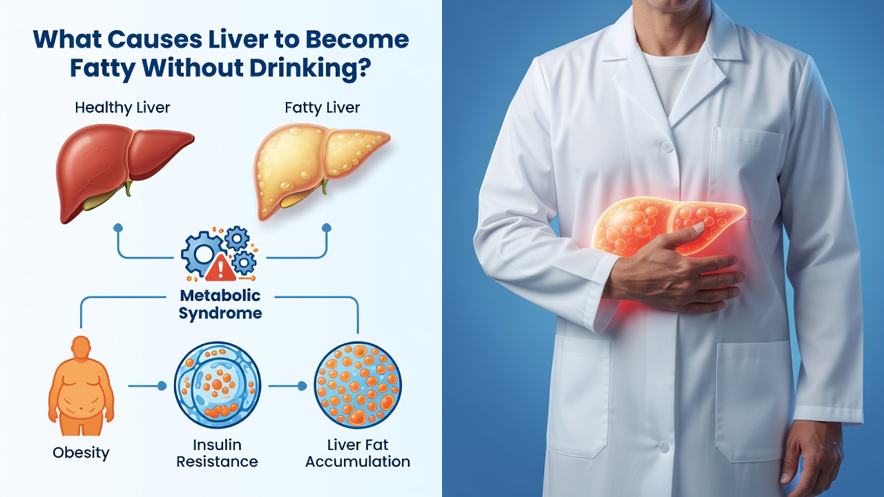
The liver becomes fatty without drinking primarily because of insulin resistance, which triggers excess fat production inside liver cells — a process called hepatic de novo lipogenesis. Obesity, type 2 diabetes, high triglycerides, and a diet heavy in refined carbohydrates and fructose are the leading drivers of this condition, clinically known as nonalcoholic fatty liver disease (NAFLD).
NAFLD — now also referred to as metabolic dysfunction-associated steatotic liver disease (MASLD) — is remarkably common. According to a 2025 epidemiological analysis published in Clinical and Molecular Hepatology, approximately 38% of all adults worldwide currently have MASLD, and that figure is projected to exceed 55% by 2040 as rates of obesity and type 2 diabetes continue to climb (PMID: 39159948). In the United States alone, experts estimate that roughly 100 million individuals carry a NAFLD diagnosis.
Understanding what causes a liver to accumulate fat without any alcohol involvement is critical for early intervention. The disease ranges from simple, asymptomatic steatosis — excess fat without inflammation — to nonalcoholic steatohepatitis (NASH), fibrosis, and ultimately cirrhosis. The good news is that the primary causes are largely metabolic and, in many cases, modifiable. For detailed guidance from a federal health authority, the National Institute of Diabetes and Digestive and Kidney Diseases (NIDDK) provides a comprehensive patient resource on NAFLD and NASH.
What Causes Liver to Become Fatty Without Drinking? The Role of Insulin Resistance
Insulin resistance is widely regarded as the central metabolic defect behind nonalcoholic fatty liver disease. When cells throughout the body stop responding normally to insulin, the pancreas compensates by producing greater amounts of the hormone. This state of chronic hyperinsulinemia has a direct effect on the liver: it dramatically upregulates de novo lipogenesis — the biochemical pathway by which liver cells (hepatocytes) synthesise new fat molecules from non-fat precursors such as glucose and fructose.
Under normal insulin signalling, the transcription factor SREBP-1c (sterol regulatory element-binding protein 1c) is activated in a controlled, meal-dependent manner to promote modest fat synthesis. In the insulin-resistant liver, however, SREBP-1c remains abnormally elevated even in the fasted state, continuously driving fatty acid production. Simultaneously, insulin resistance impairs the liver's ability to oxidise or export these newly synthesised lipids, leading to net fat accumulation — steatosis — within hepatocytes.
A landmark 2021 review published in The Lancet confirmed that NAFLD has a global prevalence of 25% and is now a leading cause of cirrhosis and hepatocellular carcinoma, with insulin resistance identified as the foundational pathogenic mechanism across patient populations (PMID: 33894145). Addressing insulin sensitivity through dietary change, physical activity, and — where appropriate — pharmacological intervention therefore remains the most evidence-supported strategy for reversing hepatic fat accumulation.
How Obesity and Visceral Fat Contribute to a Fatty Liver
Excess body weight — and specifically the accumulation of visceral adipose tissue around the abdominal organs — is one of the strongest independent risk factors for NAFLD. Visceral fat is metabolically active in a harmful way: it releases large quantities of free fatty acids (FFAs) directly into the portal circulation, which feeds straight into the liver. This constant influx of FFAs overwhelms the liver's capacity for beta-oxidation (fat burning) and VLDL-mediated export, resulting in lipid accumulation within liver cells.
The clinical numbers are striking. Research indicates that NAFLD is present in up to 75% of people who are overweight and in more than 90% of people who have severe obesity. A Mayo Clinic-affiliated study published in Clinical Gastroenterology and Hepatology established that NAFLD, already the most common liver disease in the United States at the time of publication, was expected to rise in parallel with national epidemics of obesity and type 2 diabetes (PMID: 15625647). That prediction has proven accurate.
Importantly, NAFLD is not exclusive to people with a high body mass index. Lean NAFLD — defined as fatty liver disease in individuals with a normal BMI — is a recognised clinical entity, particularly in certain Asian populations, underscoring that visceral adiposity rather than overall body weight is the more critical driver.
Why Type 2 Diabetes and Prediabetes Dramatically Increase Fatty Liver Risk
Type 2 diabetes and prediabetes share insulin resistance as their defining feature, which explains why they are so tightly linked to hepatic fat accumulation. Studies consistently find that between one-third and two-thirds of people with type 2 diabetes have NAFLD, making it the most prevalent liver condition in this population. The relationship is bidirectional: NAFLD can worsen insulin resistance, and worsening insulin resistance accelerates hepatic fat deposition — a damaging cycle.
Elevated blood glucose levels in poorly controlled diabetes provide additional substrate for de novo lipogenesis. When circulating glucose is chronically high, liver cells convert the excess glucose into acetyl-CoA, which feeds into the fatty acid synthesis pathway. Elevated insulin levels simultaneously suppress lipolysis regulation and inhibit the liver's ability to package and export triglycerides as VLDL particles, trapping fat within hepatocytes. Prediabetes — defined by impaired fasting glucose or impaired glucose tolerance — carries a comparable risk profile to full type 2 diabetes with respect to liver fat accumulation.
"NAFLD is frequently associated with insulin resistance and can be expected to increase in prevalence and severity in parallel with national epidemics of obesity and type 2 diabetes."
— Charlton MR et al., Clinical Gastroenterology and Hepatology, 2004 (PMID: 15625647)
How Diet — Especially Fructose and Refined Carbohydrates — Causes Fatty Liver Without Alcohol
Dietary composition is a powerful and often underappreciated driver of hepatic fat accumulation. Researchers have identified diets high in fructose as particularly harmful to the liver. Unlike glucose, which is metabolised by cells throughout the body, fructose is almost entirely metabolised by the liver. High fructose intake — primarily from added sugars, high-fructose corn syrup, and sugar-sweetened beverages — rapidly floods hepatocytes with substrate for de novo lipogenesis. Fructose also bypasses the key regulatory enzyme phosphofructokinase, meaning it enters the glycolytic pathway without the normal braking mechanisms that limit glucose-derived fat synthesis.
Refined carbohydrates with a high glycaemic index — white bread, white rice, pastries, and processed snack foods — produce rapid glucose spikes that provoke exaggerated insulin responses, replicating the same lipogenic signalling cascade as frank insulin resistance. Diets low in fibre and high in saturated fat also contribute by promoting gut microbiome imbalance, which independently worsens liver inflammation. Conversely, dietary patterns that emphasise whole grains, vegetables, legumes, and healthy fats — such as the Mediterranean diet — are associated with reduced hepatic fat in multiple clinical trials.
The 2023 AASLD Practice Guidance, published in Hepatology, formally recognises dietary quality as a cornerstone of NAFLD management, recommending reduced intake of fructose, saturated fat, and ultra-processed foods alongside increased physical activity (PMID: 36727674).
The Role of Gut Microbiome Imbalance and Genetics in Non-Alcoholic Fatty Liver
Two emerging areas of NAFLD research — the gut microbiome and genetic susceptibility — help explain why not everyone with obesity or insulin resistance develops fatty liver disease to the same degree. The gut microbiome, comprising trillions of bacteria in the digestive tract, communicates directly with the liver via the portal vein. Dysbiosis — an imbalance in microbial populations — increases intestinal permeability, allowing bacterial endotoxins such as lipopolysaccharide (LPS) to reach the liver and activate Toll-like receptor 4 (TLR4) signalling on Kupffer cells. This triggers the release of pro-inflammatory cytokines including TNF-α, IL-6, and IL-1β, which drive hepatic inflammation and accelerate progression from simple steatosis to NASH and fibrosis.
On the genetic side, certain gene variants substantially alter an individual's susceptibility to NAFLD. The most well-characterised is the PNPLA3 I148M variant (rs738409), which encodes a mutated form of adiponutrin — a liver enzyme involved in lipid remodelling. Individuals who carry this variant have significantly impaired hepatic lipid export and are at higher risk for both NAFLD and its progression to cirrhosis. The TM6SF2 E167K variant and MBOAT7 polymorphisms similarly affect hepatic lipid metabolism. These genetic factors help explain the higher prevalence of NAFLD observed in Hispanic populations and its relatively lower prevalence in Black Americans despite comparable rates of obesity.
Other Metabolic Conditions That Cause a Fatty Liver Without Alcohol
Beyond insulin resistance and obesity, several additional metabolic conditions create an environment in which hepatic fat accumulation becomes likely. High triglycerides (hypertriglyceridaemia) are both a consequence and a cause of NAFLD: elevated circulating triglycerides increase the substrate load delivered to the liver, while impaired VLDL export from a fatty liver further raises serum triglyceride levels. High LDL cholesterol and low HDL cholesterol — the pattern typically seen in metabolic syndrome — compound hepatic lipid stress by disrupting cholesterol homeostasis within hepatocytes.
Hypertension is independently associated with NAFLD and shares insulin resistance as a common upstream cause. Polycystic ovary syndrome (PCOS) — which involves insulin resistance and androgen excess — carries a significantly elevated NAFLD risk in women. Hypothyroidism slows hepatic fatty acid oxidation by reducing thyroid hormone-mediated mitochondrial activity, promoting fat retention in liver cells. Obstructive sleep apnoea causes intermittent hypoxia that generates oxidative stress and activates inflammatory pathways in the liver, worsening steatohepatitis independently of body weight.
Certain medications can also cause secondary fatty liver in non-drinkers. These include long-term corticosteroids (which promote visceral adiposity and insulin resistance), amiodarone (which disrupts mitochondrial beta-oxidation), methotrexate, and tamoxifen. Rapid weight loss — paradoxically — can also precipitate hepatic fat accumulation by flooding the liver with mobilised free fatty acids from peripheral fat stores.
A globally comprehensive analysis published in Hepatology found that the overall global prevalence of NAFLD rose by more than 50% between 1990–2006 and 2016–2019, reaching 38% by the later period — a trajectory driven by the simultaneous rise of these overlapping metabolic risk factors across populations worldwide.
Symptoms, Diagnosis, and What Actually Helps Reverse Fatty Liver
NAFLD is notorious for being clinically silent. The majority of people with hepatic steatosis experience no symptoms whatsoever, and some individuals do not develop symptoms even after progressing to cirrhosis secondary to NASH. When symptoms do occur, they are typically non-specific: persistent fatigue and a dull aching discomfort in the upper right quadrant of the abdomen. Elevated liver enzymes (ALT and AST) on routine blood work are often the first clinical clue, though normal liver enzymes do not exclude NAFLD.
Diagnosis is confirmed through liver ultrasound, which can detect steatosis when fat comprises more than approximately 20–30% of liver volume. More sensitive quantitative methods include MRI-based proton density fat fraction (MRI-PDFF) and controlled attenuation parameter (CAP) measured by FibroScan. Liver biopsy remains the gold standard for staging fibrosis and distinguishing simple steatosis from NASH, though it carries procedural risks and is not used as a routine screening tool.
Treatment centres on addressing the underlying metabolic drivers. Clinically meaningful weight management — including weight loss of 7–10% of body weight through dietary modification and exercise — is the most evidence-supported intervention for reducing liver fat, inflammation, and fibrosis. The Mediterranean dietary pattern is specifically endorsed by AASLD guidance. For patients diagnosed with metabolic-associated steatohepatitis (MASH) and stage 2 or 3 fibrosis, the FDA approved resmetirom (Rezdiffra) in 2024 — the first pharmacological treatment indicated specifically to reduce hepatic fat in this population. GLP-1 receptor agonists such as semaglutide also show promising hepatic benefits through their weight-reducing and insulin-sensitising effects. For authoritative treatment information, the Mayo Clinic's NAFLD treatment overview provides clinically reviewed guidance for patients and caregivers.
Evidence Summary
A sustained weight loss of 7–10% of total body weight is associated with measurable reductions in hepatic steatosis, lobular inflammation, and fibrosis scores in patients with NAFLD and NASH.
Frequently Asked Questions
Scientific References
Editorial Information
Sarah Mitchell writes educational content covering health, nutrition, and wellness topics. Her work is intended to present information in a clear and accessible format while encouraging readers to consult qualified healthcare professionals for personal medical advice.