Why Vision Deteriorates Faster After Age 50: Key Causes

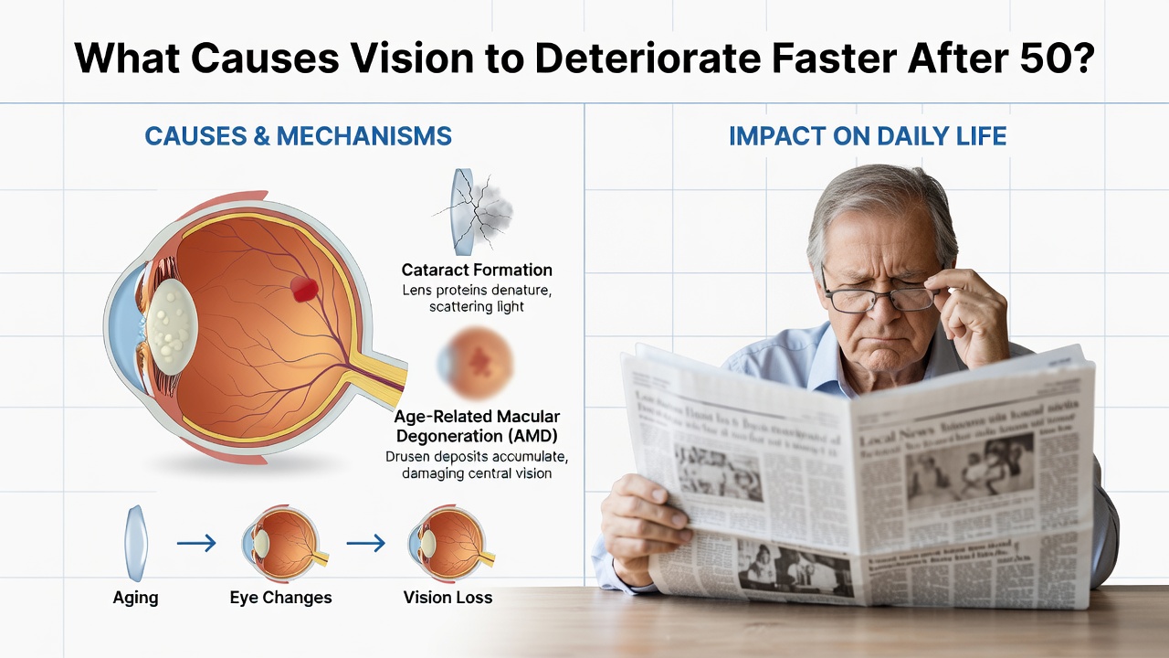
After age 50, many people notice their eyesight is not as sharp as it once was. Reading becomes harder, colors appear duller, and night driving feels riskier. This accelerated vision decline is not inevitable aging—it results from specific physiological changes in the eye's structure and biochemistry. Understanding these causes empowers you to take preventive action and maintain visual quality into your later years.
The Hardening Lens and Presbyopia
The most universal vision problem after 50 is presbyopia—difficulty focusing on close objects. This occurs because the eye's lens loses flexibility with age. The lens is made of proteins that progressively cross-link and harden over decades, reducing its ability to change shape and accommodate focus at different distances.
By age 50, nearly everyone experiences presbyopia to some degree. The ciliary muscles surrounding the lens also weaken, further diminishing focusing power. This is why bifocals or progressive lenses become necessary, and why small print becomes increasingly difficult to read without corrective aids.
Retinal Degeneration and Oxidative Stress
The retina—the light-sensitive tissue at the back of the eye—undergoes significant changes after 50. Age-related macular degeneration (AMD) is the leading cause of severe vision loss in older adults. In AMD, the macula (the central region of the retina responsible for sharp vision) gradually deteriorates due to accumulation of drusen (yellowish deposits) and oxidative damage.
Oxidative stress plays a critical role in retinal aging. The retina has exceptionally high metabolic activity and oxygen consumption, making it vulnerable to free radical damage. A foundational study demonstrated that antioxidant status directly correlates with retinal health preservation in aging populations. Cellular antioxidants like lutein, zeaxanthin, and vitamins C and E decline naturally with age, leaving the retina less protected.
Cataract Formation
Cataracts—clouding of the lens—become increasingly common after age 50. They develop when lens proteins denature and aggregate, scattering light before it reaches the retina. This results in blurred vision, reduced contrast sensitivity, and glare sensitivity, especially when driving at night.
Cataracts are accelerated by cumulative ultraviolet (UV) exposure, oxidative stress, and metabolic factors such as elevated blood sugar. People with uncontrolled diabetes show accelerated cataract development, as high glucose levels cause glycation of lens proteins, speeding protein cross-linking and clouding.
Pupil Size Reduction and Light Sensitivity Changes
After 50, the pupils become smaller and less responsive to light changes. The maximum pupil diameter in a 60-year-old is roughly half that of a 20-year-old. This dramatic reduction decreases the amount of light reaching the retina, making vision dimmer in low-light conditions and significantly impairing night vision and reading ability.
Additionally, the iris loses muscle tone and elasticity. The pupil becomes slower to adapt when moving between bright and dark environments, causing temporary vision disruption—a phenomenon familiar to anyone over 50 who enters a movie theater or dimly lit restaurant.
Vitreous Deterioration and Floaters
The vitreous humor—the clear gel filling the eye's interior—undergoes structural breakdown with age. Collagen and hyaluronic acid molecules fragment and clump, creating floaters (dark specks or strands drifting across your vision). While often harmless, floaters become more common and bothersome after 50.
More importantly, vitreous degeneration increases the risk of posterior vitreous detachment (PVD) and retinal tears or detachment. The thinning vitreous pulls away from the retina, potentially causing sudden vision loss if not promptly treated.
Corneal Flattening and Tear Film Instability
The cornea—the eye's clear front surface—flattens slightly with age, contributing to presbyopia and reduced visual acuity. Simultaneously, tear production and tear quality decline significantly after 50, leading to dry eye syndrome. The meibomian glands (which secrete lipids into tears) become less productive, destabilizing the tear film.
Chronic dry eyes blur vision and increase light scatter, compounding presbyopia and reducing overall visual clarity. This is particularly pronounced in women after menopause, when hormonal changes further reduce tear secretion.
Vascular Changes and Blood Flow Reduction
The delicate blood vessels supplying the retina and optic nerve deteriorate with age and in response to systemic vascular disease. Reduced blood flow to the eye limits oxygen and nutrient delivery to photoreceptors and supporting cells, accelerating cellular dysfunction and vision loss.
Conditions like high blood pressure and atherosclerosis directly compromise retinal circulation. Diabetic retinopathy—vessel damage from elevated blood sugar—is a leading preventable cause of vision loss in people over 50.
Mitochondrial Dysfunction
Mitochondrial health declines with age across all tissues, including the eye. Photoreceptor cells and retinal pigment epithelium cells are energy-intensive and depend heavily on mitochondrial ATP production. age-related mitochondrial DNA mutations and reduced enzyme activity compromise cellular energy metabolism, leading to photoreceptor death and vision decline.
This metabolic decline is compounded by accumulation of cellular waste products and reduced autophagy (cellular cleanup). The combination accelerates retinal aging and increases vulnerability to AMD and other degenerative conditions.
Cumulative Lifetime UV and Environmental Damage
Years of sun exposure without adequate UV protection accumulate in eye tissues. Ultraviolet radiation triggers oxidative stress and accelerates lens protein cross-linking, advancing cataract formation. By age 50, most people have received substantial cumulative UV exposure.
Environmental pollutants and smoke exposure similarly generate oxidative stress in ocular tissues, amplifying the aging process. This explains why smokers and individuals with high lifetime sun exposure experience vision loss earlier and more severely than protected counterparts.
Evidence-Based Prevention and Management
While some age-related vision changes are unavoidable, research supports several protective strategies. UV protection through quality sunglasses and hats reduces cataract and AMD risk. The Age-Related Eye Disease Study (AREDS) confirmed that antioxidant supplementation—including vitamins C and E, zinc, lutein, and zeaxanthin—slows progression of advanced AMD by approximately 25%.
Maintaining stable blood sugar, managing blood pressure, and avoiding smoking are critical. Regular comprehensive eye exams after 50 enable early detection of glaucoma, AMD, and diabetic retinopathy—conditions where early intervention preserves vision. Adequate sleep supports retinal health and tear production. Screen time breaks using the 20-20-20 rule (every 20 minutes, look at something 20 feet away for 20 seconds) reduce digital eye strain and temporary accommodation fatigue.
Frequently Asked Questions
Scientific References
Editorial Information
Sarah Mitchell writes educational content covering health, nutrition, and wellness topics. Her work is intended to present information in a clear and accessible format while encouraging readers to consult qualified healthcare professionals for personal medical advice.